November 2019 | Volume 21 No. 1
Digital Dentistry
“Dentists will still be crucial for diagnosis and selecting the best treatments,” said Dr Walter Yu-hang Lam, Clinical Assistant Professor in Prosthodontics, “but the basics will be done efficiently and economically by robots.
“There have already been many digital developments in other areas. Dentists have started to use optical scanners (intra-oral scanner or IOS) to scan patients’ teeth and create 3D models for diagnosis and treatment of disease. They are also scanning and ‘digitising’ patient data for the design of dental restorations via computer, and many novel tooth-coloured dental materials – such as zirconia – are now fabricated by the CAD/CAM (computerised aided design and manufacturing) process, replacing ugly metal restorations.”These advances are not only improving the accuracy and efficacy of treatments for patients, they are also more durable and save dentists’ time – both of which bring monetary savings, enabling cheaper treatments for the public in the future.
Most of a dentist’s clinical time is spent replacing patients’ missing teeth or inserting fillings, and one of the prime requirements for replacing or filling teeth is harmony to the patient’s masticatory system.
“Traditionally, a mechanical device called an articulator is used to simulate patients’ jaw movement,” said Dr Lam. “However, articulators are expensive and because they are mechanical devices and cannot accommodate the wide range of variations in the human system – for example most patients’ faces are not symmetrical – so simulation precision is limited and usually the dentist has to do ‘chair-side adjustments’ in the patients’ mouth.”
His team has introduced a clinical technique for a virtual articulator mounting with a 3D facial camera (stereophotogrammetry) to enable a simulation function that is far more precise and can account for patient’s individual variations. Moreover, with the exception of the 3D camera, the equipment used is modified from things readily available in the dental clinic.
“Accurate articulator-mounted casts are essential for occlusion [chewing] analysis and for fabrication of dental restorations. In the past there have been problems with getting the head position of the patient [known as the natural head position or NHP] marked accurately,” said Dr Lam.
He explained that his new technique uses the 3D camera to define the horizontal plane in the capture area using a reference board (calibration) whose position is defined by vertical/horizontal light beams coupled with a spirit level normally used for building surveying. The horizontal plane as designated by the reference board can be transferred to the subsequent 3D photo of patients in NHP.
Virtual patient models
The team has also made advances in the development of better virtual patient models (VPM), which are both functional and can aesthetically simulate real patients, for CAD/CAM design of tooth restoration.
Designs include combining 3D teeth to a 3D face to create a two-part ‘facebow’. Dr Lam said: “Teeth and face are captured by two imaging devices – an intraoral scanner and a 3D camera. We proposed a non-invasive method to combine these two images together using a two-part virtual facebow.
“Unlike other researchers who propose using a tailor-made intraoral device, we modified the readily available dental impression trays originally used for making teeth impressions in any dental clinic. We assembled two trays together using two Lego bricks to relate teeth to the face in the 3D camera.
“Our virtual facebow facilitates the radiation-free registration of the teeth to a face image for transfer to a virtual articulator. The facebow is easy to make with minimal materials and adjusts to fit different people. Most importantly, its error in tooth registration is less than one millimetre.”
Dr Lam and his team have worked extensively with Dr Wang Zheng from the Department of Mechanical Engineering to develop robots for dental treatment. In particular he is interested in how to enable virtual dental patients and dental robots to work together so as to reduce dentist’s input time, which will have the knock-on effect of making dental treatment more affordable to the community.
Together, they have developed a robotic system for assisting dental drilling procedures, specifically designed to work in the smaller workspace – the mouth – in which dentists work. The result is a soft-bracing actuator which has improved the system stiffness to 10 times higher than non-braced models.
Their aim in developing the system was to relieve the burden on dentists, improve the efficiency of dental procedures and reduce the possibility of human error during treatment.
“We hope the robotic system we have proposed will accelerate automation in dental treatments,” said Dr Lam. “With several iterations of the design and algorithm, we also hope clinical trials can be conducted in the near future. The development of virtual patient models and dental robots will facilitate dentists to provide treatment to patients who live in remote areas or have limited accessibility.”
Up next, Dr Lam will work with researchers at the University of Leeds, led by Professor Andrew Keeling, to use mobile phones to digitalise dental patients (VPM) and identify oral diseases via photography. “You could say that the ultimate aim is to achieve a ‘patient selfie’,” he explained. “Then the AI can tell if the patient has an oral disease.”
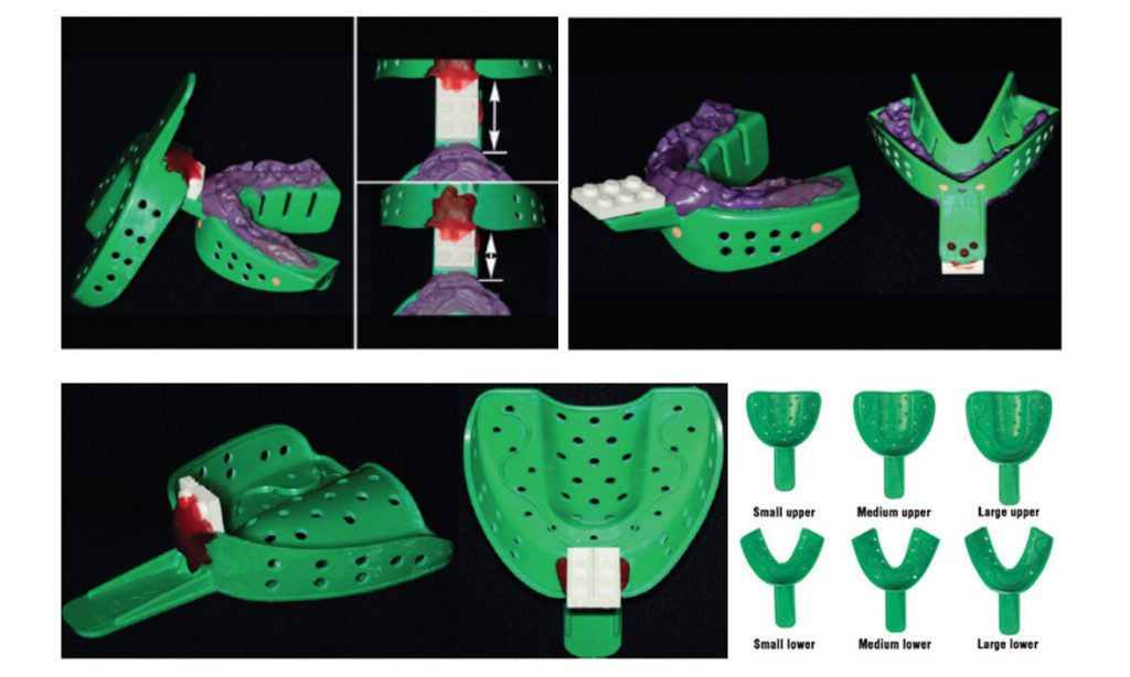
Assemble of virtual facebow for individual patients.
The development of virtual patient models and dental robots will facilitate dentists to provide treatment to patients who live in remote areas or have limited accessibility.

DR WALTER YU-HANG LAM
